Disease Evolutionary Ecology
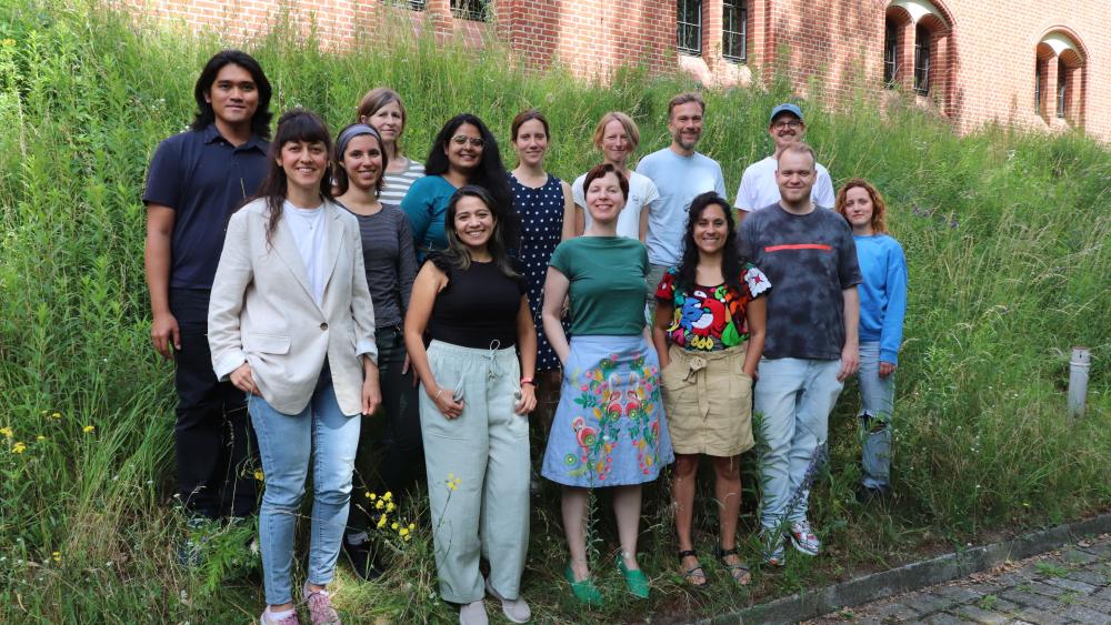
Disease Evolutionary Ecology Group, June 2024
Back row (left to right): Vanderville Villegas (Andy), Athena Karapli-Petritsopoulou, Dagmar Frisch, Amruta Rajarajan, Pauline Thomé, Justyna Wolinska, Jürgen Strassert, Fabian Flücken.
Front row (left to right): Zeynep Daldaban, Erika Martinez Ruiz, Elisabeth Funke, Kristel Sanchez, Benjamin Schupp, Sarah Lauck.
We study evolutionary and ecological processes mediated by parasitism in aquatic ecosystems. Parasites are ubiquitous and impose strong selection on their hosts to evolve resistance, while themselves being under strong selection to undermine host defenses. We aim to improve understanding of the interface between host-parasite co-evolution and major ecological processes, under the influence of global environmental change. We focus on questions such as how parasitism contributes to the maintenance of genetic diversity, how it shapes interactions between host and non-host species, and how anticipated environmental challenges will modulate the occurrence of disease.
Our group uses several empirical approaches. Through field studies of natural populations, we are exploring the links between coevolution and genetic diversity, as well as between environmental components and occurrence of disease. Our field sites consist of sets of large permanent lakes, representing a gradient of environmental conditions. Specifically, we track genetic changes in populations and communities over both time and space (employing molecular approaches, e.g. next-generation-sequencing). Another component of our research involves laboratory experiments (single-generation experiments, as well as long-term surveys in microcosms). As a main model organism, we have concentrated on the crustacean Daphnia, which is well-established in ecological, evolutionary and genomic research, and is a key component of aquatic food webs. In addition, we study various phytoplankton species and their natural fungal (chytrid) parasites.
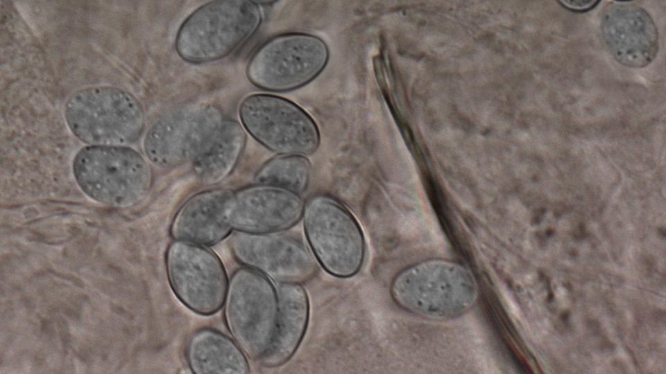
Spores of Caullerya mesnili. | Image: Petr Jan Juracka
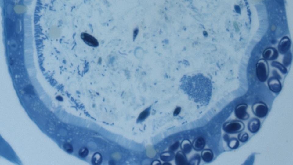
Spore clusters of Caullerya mesnili in the Daphnia gut; histological section. | Image: Wolinska Lab
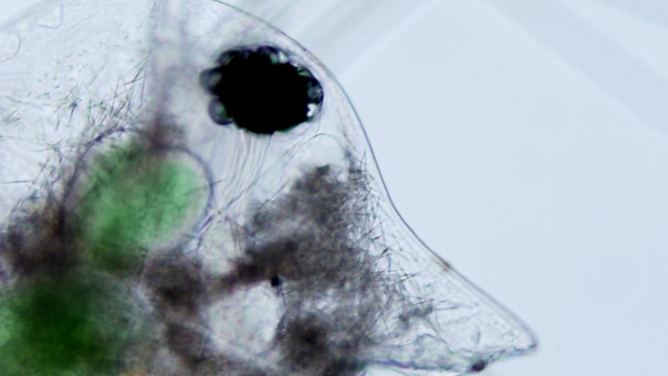
Daphnia infected with yeast parasite, Metschnikowia sp. | Image: Wolinska Lab
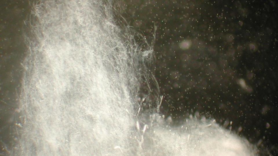
Sporulating water mould - parasite of Daphnia. | Image: Wolinska Lab
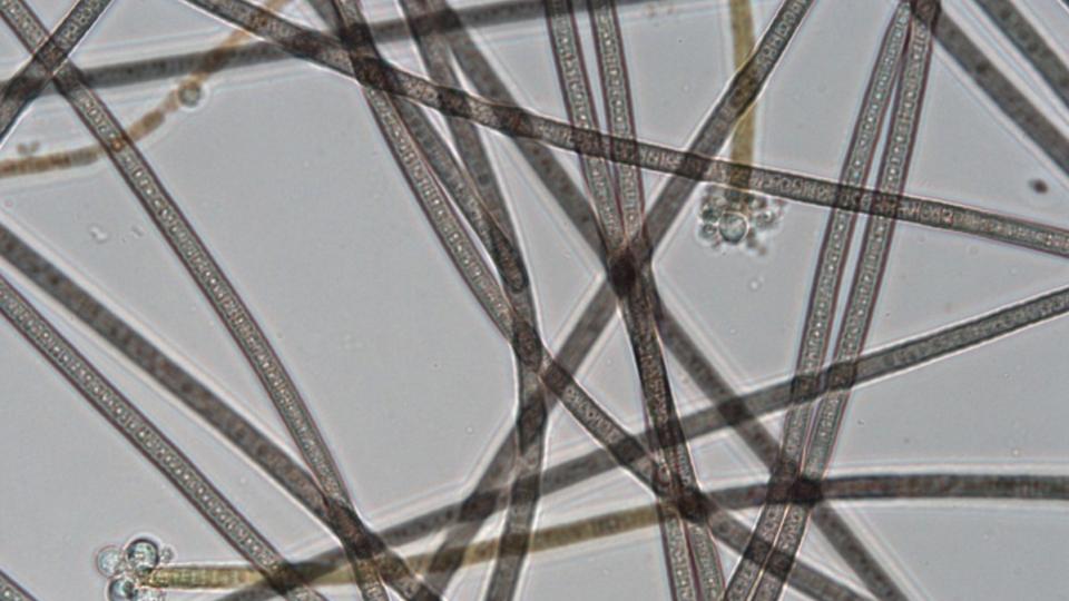
Cyanobacterium Planktothrix rubescens infected by chytrid fungus Rhyzophydium megarrhizum. Spherical structures are chytrid sporangia. | Image: Ramsy Agha / IGB
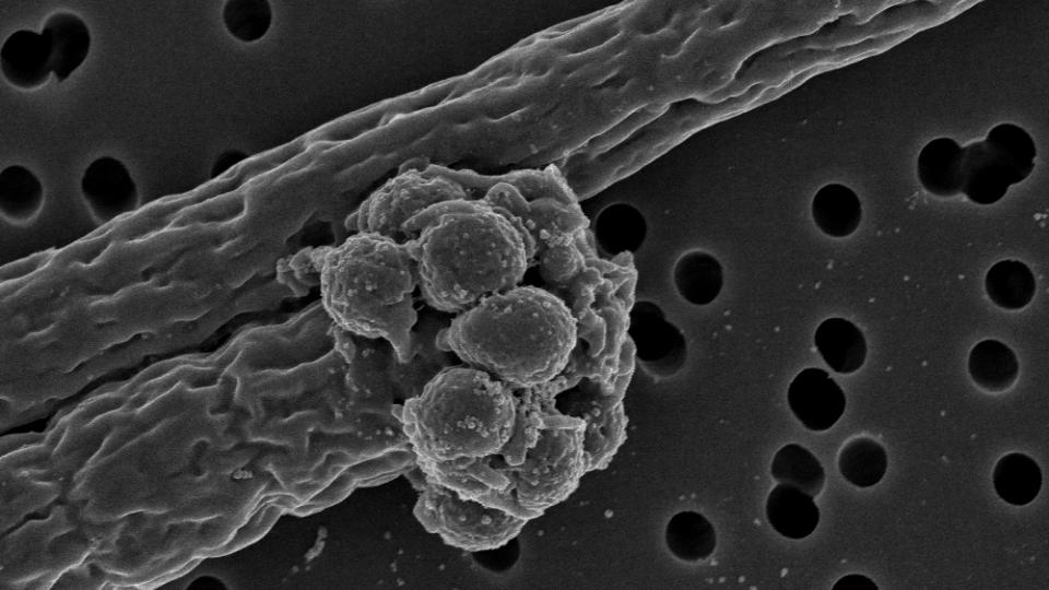
Chytrid Rhyzophydium megarrhizum encysted on a filament of its host, the cyanobacterium Planktothrix agardhii. | Image: Ramsy Agha / IGB
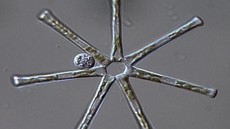
Chytrid on Asterionella formosa. | Image: Melanie Gerphagnon
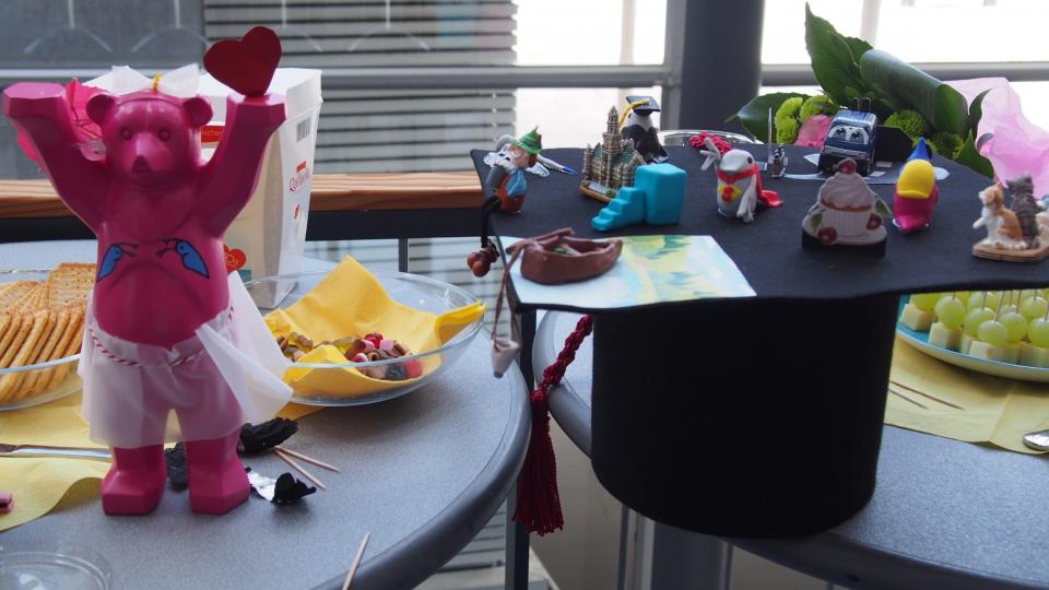
Daphnia everywhere; PhD defense of Johanna. | Photo: Wolinska Lab
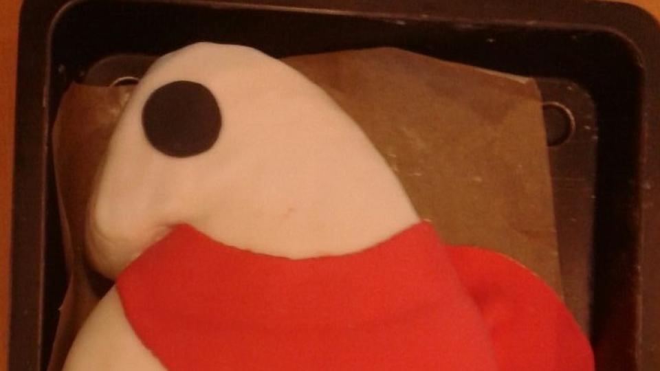
Super Daphnia (project of Johanna & Manja). | Photo: Wolinska Lab
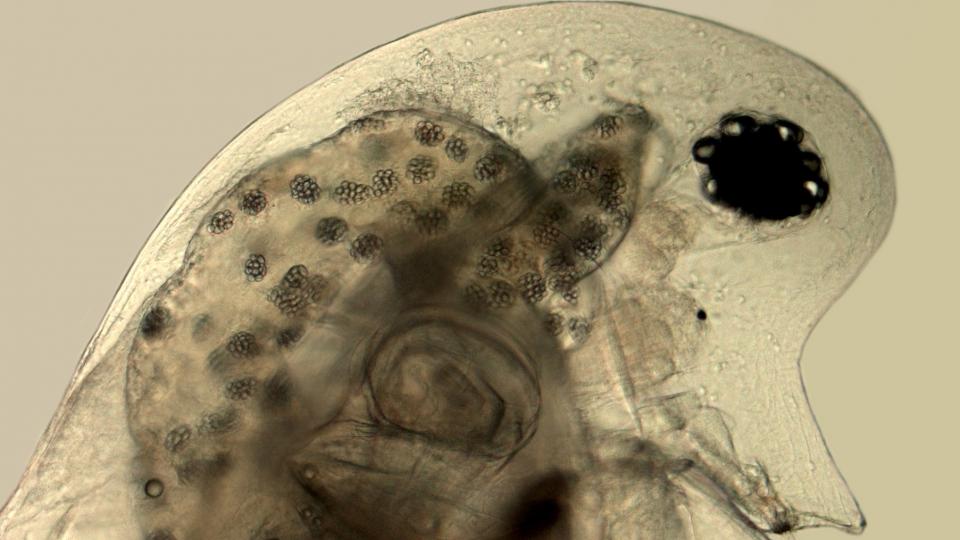
Daphnia galeata infected with a gut parasite Caullerya mesnili; spore clusters visible in the gut epithelium. | Image: Petr Jan Juracka
Publications 2016. | Image: Wolinska Lab